The skull, also known as the cranium, typically constitutes a bony encasement for the brain in vertebrates. In certain fish and amphibians, this structure is cartilaginous. Positioned at the anterior extremity of the vertebrate body, the skull serves as the primary cephalic structure.
The skull, or cranium, is typically a bony enclosure around the brain of a vertebrate. In some fish and amphibians, the skull is of cartilage. The skull is at the head end of the vertebrate.
The human skull is composed of two principal components: the neurocranium and the facial skeleton, the latter having evolved from the first pharyngeal arch. This cranial structure represents the most anterior segment of the axial skeleton, resulting from cephalization and the encephalic vesicular enlargement. It integrates various specialized sensory organs, including the eyes, ears, nose, and tongue, and in fish, specialized tactile structures like barbels situated near the oral cavity.
The skull is constituted by three categories of bones—cranial bones, facial bones, and ossicles—and is formed from numerous fused flat and irregular bone elements. The cranial bones are interconnected by robust fibrous junctions, termed sutures, and feature numerous foramina, fossae, processes, and sinuses. In zoological contexts, cranial apertures are referred to as fenestrae; the most conspicuous of these is the foramen magnum, through which the brainstem extends to connect with the spinal cord.
Within human anatomy, the neurocranium, also known as the braincase, is subdivided into the calvaria and the endocranium, which collectively form the cranial cavity enclosing the brain. The internal periosteum contributes to the dura mater, while the facial skeleton, or splanchnocranium, includes the mandible as its largest bone. The mandible articulates with the temporal bones of the neurocranium via the paired temporomandibular joints. The skull itself establishes articulation with the vertebral column at the atlanto-occipital joint. Complete development of the human skull typically occurs two years post-natally.
The skull serves multiple functions, encompassing the physical protection of the brain, the provision of attachment points for cervical, facial, and masticatory muscles, and the formation of stable orbital sockets and external auditory structures (ear canals and auricles) crucial for stereoscopic vision and sound localization. Furthermore, it defines the nasal and oral cavities, facilitating enhanced olfaction, gustation, and digestion, and contributes to phonation through acoustic resonance within its various cavities and sinuses. In certain animal species, such as ungulates and elephants, the skull additionally plays a role in anti-predator defense and sexual selection by providing the structural basis for horns, antlers, and tusks.
The etymology of the English term skull likely traces to the Old Norse word skulle. Concurrently, the Latin term cranium originates from the Greek root κρανίον (kranion).
Structure
Humans
The human skull constitutes the bony framework of the head within the human skeleton. It provides support for facial structures and creates a protective cavity for the brain. Analogous to the crania of other vertebrates, it safeguards the brain from potential trauma.
The skull is composed of three embryologically distinct components: the neurocranium, the sutures, and the facial skeleton. The neurocranium, also termed the braincase, establishes the protective cranial cavity that envelops and contains the brain and brainstem. The superior regions of the cranial bones collectively constitute the calvaria, or skullcap. The facial skeleton, also known as the membranous viscerocranium, comprises the bones that support the face and incorporates the mandible.
The cranial bones are interconnected by fibrous joints, specifically sutures, which are synarthrodial (immovable) articulations formed through bony ossification, with Sharpey's fibers allowing for a degree of flexibility. Occasionally, accessory bone fragments, referred to as Wormian bones or sutural bones, may be present within these sutures. These are most frequently observed along the trajectory of the lambdoid suture.
Bones
The human skull is generally understood to comprise 22 distinct bones: eight cranial bones and fourteen facial skeleton bones. Within the neurocranium, these include the occipital bone, two temporal bones, two parietal bones, the sphenoid bone, the ethmoid bone, and the frontal bone.
The fourteen bones of the facial skeleton consist of the vomer, two inferior nasal conchae, two nasal bones, two maxillae, the mandible, two palatine bones, two zygomatic bones, and two lacrimal bones. While some anatomical sources may enumerate paired bones as a single entity, or consider the maxilla as two distinct parts, and others may incorporate the hyoid bone or the three middle ear ossicles (malleus, incus, and stapes), the prevailing consensus regarding the total number of bones in the human skull remains twenty-two.
Among these bones, the occipital, parietal, and frontal bones of the neurocranium, along with the nasal, lacrimal, and vomer bones of the facial skeleton, are classified as flat bones.
Cranial Cavities and Foramina
The skull incorporates paranasal sinuses, which are air-filled cavities, alongside numerous foramina. These sinuses are internally lined with respiratory epithelium. Their established physiological roles include reducing the overall weight of the skull, enhancing vocal resonance, and conditioning inhaled air by warming and moistening it before it reaches the nasal cavity.
Foramina represent various apertures within the skull. The most prominent among these is the foramen magnum, located in the occipital bone, which facilitates the transit of the spinal cord, cranial nerves, and associated blood vessels.
Cranial Processes
The skull features multiple anatomical projections, notably including the mastoid process and the zygomatic processes.
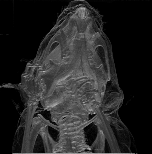
Vertebrate Skull Morphology
Cranial Fenestrae
Skeletal Components
The jugal bone is a cranial element prevalent across most reptiles, amphibians, and avian species. In mammalian anatomy, this bone is frequently referred to as the zygomatic bone or malar bone.
The prefrontal bone serves as a distinct skeletal component, articulating between the lacrimal and frontal bones in numerous tetrapod crania.
Ichthyological Cranial Structures
Fish skulls are typically composed of a series of loosely articulated bones. Lampreys and sharks, for instance, exhibit a cartilaginous endocranium, where both the upper and lower jaws exist as distinct, separate components. In contrast, bony fishes possess additional dermal bone, which contributes to a more integrated skull roof, particularly evident in lungfish and holost fish. The lower jaw fundamentally shapes the chin region.
A more rudimentary cranial architecture characterizes jawless fish, where the cranium typically manifests as a trough-shaped cartilaginous framework. This structure only partially encases the brain and is associated with capsules for the inner ears and a singular nostril. A defining characteristic of these fish is the complete absence of jaws.
Cartilaginous fish, exemplified by sharks and rays, also exhibit relatively simple, and presumably ancestral, skull configurations. Their cranium forms a singular, protective enclosure around the brain, encompassing its ventral and lateral aspects, yet consistently featuring at least a partial dorsal opening, often a substantial fontanelle. The most anterior cranial region comprises a cartilaginous rostral plate and capsules that house the olfactory organs. Posterior to these are the orbits, followed by another pair of capsules encasing the inner ear structures. Towards the posterior, the skull gradually narrows, culminating in the foramen magnum, situated directly superior to a solitary condyle that articulates with the first vertebra. Furthermore, the cranium is punctuated by smaller foramina at various locations, facilitating the passage of cranial nerves. The jaws themselves are composed of distinct cartilaginous arches, almost invariably separate from the main cranial structure.
Ray-finned fish demonstrate substantial evolutionary divergence from the ancestral cranial blueprint. Their skull roof is typically well-developed; although the precise homologous relationships of its bones to those of tetrapods remain ambiguous, they are conventionally assigned analogous nomenclature for descriptive ease. Conversely, other cranial components may exhibit reduction; for instance, the post-orbital cheek region is often diminished, and osseous material between the enlarged orbits is minimal or absent. The upper jaw frequently derives predominantly from the premaxilla, with the maxilla positioned more posteriorly, and an accessory bone, the symplectic, serving to connect the jaw apparatus to the remainder of the cranium.
While the crania of fossil lobe-finned fish bear morphological resemblances to those of early tetrapods, this congruence does not extend to extant lungfishes. In living lungfishes, the skull roof is incompletely ossified, comprising numerous, somewhat irregularly configured bones that lack direct homologous relationships with tetrapod cranial elements. The upper jaw is exclusively constituted by the pterygoids and vomers, all of which are dentigerous. A significant proportion of the skull remains cartilaginous, contributing to an overall reduction in its structural complexity.
Tetrapod Cranial Anatomy
The crania of early tetrapods bore a strong resemblance to those of their lobe-finned fish ancestors. The skull roof comprises a sequence of plate-like bones, such as the maxilla, frontals, parietals, and lacrimals, among others. This structure overlies the endocranium, which is analogous to the cartilaginous skull found in sharks and rays. Components that form the human temporal bone are also integrated into this skull roof series. Additionally, a plate consisting of four pairs of bones, including the vomer and palatine bones, constitutes the roof of the oral cavity. The cranial base is constructed from several bones, many of which exhibit homology with elements of the sphenoid and occipital bones in mammals. Lastly, the lower jaw is multi-boned, with only its most anterior component, the dentary, being homologous to the mammalian mandible.
Among extant tetrapods, numerous ancestral bones have either undergone reduction, complete loss, or fusion into diverse configurations.
Avian Skulls
Avian skulls are characterized by a diapsid structure, similar to that observed in reptiles, and often feature a prelacrimal fossa, which is also present in some reptilian species. A singular occipital condyle is a distinguishing feature. The avian skull is primarily composed of five significant bones: the frontal bone, forming the cranial vault; the parietal bone, located at the posterior aspect of the cranium; the premaxillary and nasal bones, which collectively form the upper beak; and the mandible, constituting the lower beak. Typically, a bird's skull accounts for approximately 1% of its total body weight. The ocular region occupies a substantial portion of the skull and is encircled by a sclerotic eye-ring, a structure comprising numerous small bones. This particular anatomical trait is also evident in reptiles.
Amphibian Skulls
Extant amphibians generally possess significantly reduced skulls, where many bones are either absent or have been entirely or partially supplanted by cartilage. Notably, in mammals and birds, cranial modifications have evolved to accommodate brain expansion. The extensive fusion among various bones is particularly pronounced in avian species, often rendering the identification of individual skeletal components challenging.
Cranial Development
The skull represents an intricate anatomical construct, with its constituent bones developing through both intramembranous and endochondral ossification processes. The bones forming the skull roof, which include elements of the facial skeleton and the lateral and superior aspects of the neurocranium, originate as dermal bones via intramembranous ossification; however, the temporal bones are an exception, forming through endochondral ossification. Conversely, the endocranium, which encompasses the bones that support the brain—specifically the occipital, sphenoid, and ethmoid bones—is predominantly formed by endochondral ossification. Consequently, the frontal and parietal bones are exclusively membranous in origin. The morphology of the skull base and its associated fossae—the anterior, middle, and posterior cranial fossae—undergoes dynamic transformations. The anterior cranial fossa, in particular, exhibits significant changes during the first trimester of gestation, a period when cranial defects frequently emerge. The prenatal development of the anterior cranial fossa is characterized by non-uniform growth. During the initial trimester, allometric growth is observed, with the longitudinal dimension expanding from 5 to 17 millimeters between the 8th and 14th weeks of fetal development. Concurrently, the angle of the anterior cranial fossa diminishes, and its depth increases in the direction of the middle cranial fossa. In the second trimester, growth persists but adopts a more uniform pattern, with only minor alterations in the angle of the anterior cranial fossa. A progressive reduction in the angle between the lesser wings of the sphenoid bone is noted as the depth of the anterior cranial fossa increases within the frontal plane.
The human skull comprises 44 distinct bony elements at birth. Throughout development, numerous these elements progressively coalesce into solid bone, exemplified by the frontal bone. Initially, the cranial vault bones are demarcated by areas of dense connective tissue known as fontanelles. These include six distinct fontanelles: one anterior (frontal), one posterior (occipital), two sphenoid (anterolateral), and two mastoid (posterolateral). At parturition, these fibrous and mobile regions facilitate both the birthing process and subsequent cranial expansion. Such growth can exert substantial tension on the "obstetrical hinge," the junction of the occipital bone's squamous and lateral components. A potential sequela of this tension is the rupture of the great cerebral vein. With continued growth and ossification, bone progressively replaces the connective tissue of the fontanelles, forming sutures. The five primary sutures are the two squamous, one coronal, one lambdoid, and one sagittal. While the posterior fontanelle typically ossifies by eight weeks postpartum, the anterior fontanelle may persist until eighteen months of age. Situated at the confluence of the frontal and parietal bones, the anterior fontanelle constitutes a palpable "soft spot" on an infant's forehead. Careful observation permits the assessment of an infant's heart rate by noting the subtle pulsations visible through the anterior fontanelle.
The neonatal skull exhibits a disproportionately large size relative to other somatic structures. The facial skeleton measures approximately one-seventh the size of the calvaria, contrasting with its adult proportion of half the size. The cranial base is characterized by its brevity and narrowness, notwithstanding the near-adult dimensions of the inner ear.
Clinical Significance
Craniosynostosis represents a pathological condition characterized by the premature fusion of one or more fibrous sutures within an infant's skull, thereby altering the cranial growth trajectory. As the skull is unable to expand perpendicularly to the fused suture, compensatory growth occurs predominantly in the parallel plane. While this compensatory growth may occasionally accommodate the developing brain, it frequently leads to an atypical cranial morphology and distinctive facial dysmorphism. Should this compensatory mechanism prove insufficient to provide adequate space for the expanding brain, craniosynostosis can precipitate elevated intracranial pressure, potentially resulting in visual deficits, sleep disturbances, feeding difficulties, or impaired cognitive development.
A "copper beaten skull" describes a phenomenon where sustained, intense intracranial pressure deforms the internal cranial surface. This nomenclature derives from the characteristic appearance of the inner skull, which resembles the indentations produced by a ball-peen hammer, a tool commonly employed by coppersmiths. This condition predominantly affects pediatric populations.
Injuries and Treatment
Cerebral injuries pose a significant threat to life. Typically, the skull safeguards the brain from trauma due to its exceptional resistance to deformation; it stands among nature's least deformable structures, requiring approximately one ton of force to reduce its diameter by one centimeter. Nevertheless, certain head injuries can lead to elevated intracranial pressure, often mediated by conditions like a subdural hematoma. Such elevated pressure can induce cerebral herniation through the foramen magnum (termed 'coning'), as the brain lacks space for expansion, potentially resulting in severe neurological damage or mortality unless immediate surgical intervention alleviates the pressure. Consequently, individuals diagnosed with concussion necessitate rigorous monitoring. Repeated concussions can induce structural changes in the cranial bones, which serve as the brain's protective covering.
Evidence suggests that a cranial surgical procedure, known as trepanning, was occasionally performed as early as the Neolithic period. This intervention entailed drilling a burr hole into the cranium. Paleopathological analysis of skulls from this era indicates that some patients survived for extended periods post-procedure. It is also posited that trepanning may have been conducted for purely ritualistic or religious purposes. Contemporary medical practice continues to utilize a similar procedure, now commonly referred to as a craniectomy.
In March 2013, researchers in the United States achieved a medical first by replacing a substantial portion of a patient's skull with a precision 3D-printed polymer implant. Approximately nine months subsequently, the inaugural complete cranial replacement using a 3D-printed plastic insert was successfully performed on a Dutch female patient. This patient had been afflicted with hyperostosis, a condition characterized by increased skull thickness and subsequent cerebral compression.
Research conducted in 2018 by Harvard Medical School investigators in Boston, with funding from the National Institutes of Health (NIH), proposed that immune cells, in conjunction with bone marrow, access inflamed brain tissue following injury not through the bloodstream, but via minute channels within the skull.
Transgender Procedures
Reconstructive surgical procedures, such as facial feminization surgery or facial masculinization surgery, involve the modification of sexually dimorphic skull features. These interventions aim to reshape and resize facial characteristics to align them more closely with those typically associated with the desired sex, constituting a significant component of gender dysphoria treatment for transgender individuals.
Society and Culture
Artificial cranial deformation represents a historical practice observed in various cultures. This process involved applying pressure to an infant's skull using cords and wooden boards, commencing shortly after birth and continuing for several years, often resulting in substantial alterations to cranial morphology.
Osteology
The skull and dentition, similar to facial features, can provide insights into an individual's life history and geographical origin. Forensic scientists and archaeologists employ both quantitative and qualitative characteristics to reconstruct the appearance of the skull's owner. In contexts where substantial skeletal remains are discovered, such as at Spitalfields in the UK or Jōmon shell mounds in Japan, osteologists analyze traits like length, height, and width proportions to ascertain the relationships between the studied population and other extant or extinct groups.
Around 1800, the German physician Franz Joseph Gall developed the theory of phrenology, which posited a correlation between specific cranial features and an individual's personality traits or intellectual capacities. This theory is currently regarded as pseudoscientific.
Sexual Dimorphism
During the mid-nineteenth century, anthropologists prioritized differentiating between male and female skulls. James McGrigor Allan, a contemporary anthropologist, asserted that the female brain resembled that of an animal, leading to the declaration that women were inherently more emotional and less rational than men. McGrigor further concluded that female brains were analogous to those of infants, thereby classifying women as inferior. To reinforce these assertions of female inferiority and suppress feminist discourse, other anthropologists engaged in studies of the female skull. These cranial measurements formed the foundation of craniology and were also utilized to establish a perceived link between women and Black individuals.
While minimal differences exist between male and female skulls in early life, adult male skulls are generally larger and more robust compared to female skulls, which are typically lighter and smaller, possessing approximately 10 percent less cranial capacity. However, subsequent research indicates that female skulls may be marginally thicker, potentially rendering males more vulnerable to head injuries. Conversely, other investigations suggest that male skulls exhibit greater thickness in specific regions. Some studies also report a higher susceptibility to concussion among females than males. Furthermore, male skulls have demonstrated a tendency to maintain density with age, potentially contributing to head injury prevention, whereas female skull density tends to decline slightly over time.
Male skulls typically exhibit more prominent supraorbital ridges, glabella, and temporal lines. In contrast, female skulls generally feature rounder orbits and narrower jaws. On average, male skulls possess larger, broader palates, squarer orbits, more substantial mastoid processes, larger sinuses, and larger occipital condyles compared to female skulls. Male mandibles are also characterized by squarer chins and thicker, rougher muscle attachments than their female counterparts.
Craniometry
The cephalic index is defined as the ratio of head width to head length (anterior-posterior), multiplied by 100. This index is also applied in the classification of animals, particularly canines and felines. Typically, the width measurement is taken inferior to the parietal eminence, while the length extends from the glabella to the occipital point.
Humans may be categorized as:
- Dolichocephalic — characterized by a long head.
- Mesaticephalic — characterized by a medium-length head.
- Brachycephalic — characterized by a short head.
The vertical cephalic index quantifies the ratio derived from multiplying the head's height by 100 and subsequently dividing by its length.
Human crania can be categorized based on their vertical cephalic index as:
- Chamaecranic: Characterized by a low skull height.
- Orthocranic: Exhibiting a medium skull height.
- Hypsicranic: Possessing a high skull height.
Related Terminology
- Chondrocranium: A primitive skeletal structure composed of cartilage.
- Endocranium
- Epicranium
- Pericranium: A membrane enveloping the external surface of the cranium.
Historical Context
Trepanning, the surgical creation of a hole in the skull, is recognized as the most ancient surgical procedure supported by archaeological evidence, including cave paintings and human skeletal remains. For instance, at a burial site in France dating to 6500 BCE, 40 of 120 prehistoric skulls exhibited trepanation holes.
References
This article integrates public domain text from page 128 of the 20th edition of Gray's Anatomy (1918).
This article, as of its current revision, incorporates content from "Morphometric evaluation of the anterior cranial fossa during the prenatal stage in humans and its clinical implications", authored by Wojciech Derkowski, Alicja Kędzia, Krzysztof Dudek, and Michał Glonek. This content is licensed for reuse under the Creative Commons Attribution-ShareAlike 4.0 International License, though not under the GFDL, and all associated terms must be observed.
- Skull Module (California State University Department of Anthology)
- Bird Skull Collection Bird skull database with very large collection of skulls (Agricultural University of Wageningen)
- Human Skulls / Anthropological Skulls / Comparison of Skulls of Vertebrates (PDF; 502 kB)